
Diagnostic ultrasound imaging in sports medicine has proven to be a reliable imaging modality in a variety of conditions that are often difficult to evaluate using x-ray, computed tomography (CT) scans, or magnetic resonance imaging (MRI). Among its many advantages is that, since it does not use ionizing radiation, it can be repeated throughout the course of treatment of an injury without concern for the total amount of radiation delivered to the patient.
Ultrasound imaging in sports medicine relies on high frequency sound waves that are transmitted to the skin overlying the area of interest such as the wrist, shoulder, or knee. As these waves encounter different tissues, some waves are reflected back to their source while others continue on until they too are reflected. At the source, which is a device known as a transducer, the returning sound waves are converted to electrical impulses that are sent to a computer which uses these impulses to construct an image of the area being evaluated.
As a rule, higher the frequency sound waves will yield more detailed images. The downside to this higher detail is that high frequency sound does not travel beyond a few centimeters in depth. Hence, ultrasound imaging in sports medicine is limited to superficial structures.
Ultrasound imaging is frequently used to diagnose injuries to the the tendons and ligaments (the rotator cuff) of the shoulder. Such injuries are difficult to diagnose using x-rays or MRI but ultrasound, because it is rapid and provides images while the patient performs certain maneuvers such as placing he arm in different positions, can detect even the smallest of injuries.
The wrist is another area that is frequently injured during athletic activities or by repetitive motion injury such as typing. Ultrasound imaging in these cases can detect tears or bruising of the tendons of the wrist and can also detect excess fluid collections in the area of the wrist known as the carpal tunnel.
Many athletic injuries involve the knee or its adjacent muscles and tendons. While ultrasound cannot penetrate bone it is ideally suited for detecting the muscle tears and bruising that usually accompany knee injuries. And, since ultrasound studies can be repeated as often as required, they are an efficient method for evaluating how such injuries are healing.
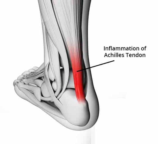
After the knee and wrist, the most frequent athletic injuries involve the ankle and its attached structures. Ultrasound is a very reliable imaging technology for evaluation of injuries to the Achilles tendon and adjacent tissues.
In addition to the specific structures already mentioned, ultrasound imaging in sports medicine is often used to detect injuries such as bruises or muscle strains or tears. Although such injuries can also be diagnosed with MRI images, ultrasound is the technology of choice because it takes less time to acquire the necessary images. Ultrasound imaging of athletic images is also more cost effective than images obtained with Ct or MRI. The choice of which imaging modality to be used will always rest with the attending physician.
In summary, ultrasound imaging in sports medicine is a valuable diagnostic tool and will remain a vital part of sports medicine for years to come.